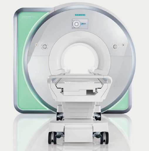
MRI
Reserve now
Service Description
Magnetic resonance imaging is a medical imaging technique used in radiology to form pictures of the anatomy and the physiological processes of the body.
Preparations
- Guidelines about eating and drinking before an MRI exam vary with the specific exam.
- The radiologist or technologist may ask if you have allergies of any kind, such as an allergy to iodine or x-ray contrast material, drugs, food, the environment, or asthma.
- The radiologist should also know if you have any serious health problems, or if you have recently had surgery.
- Women should always inform their physician or technologist if there is any possibility that they are pregnant.
- If you have claustrophobia (fear of enclosed spaces) or anxiety, you may want to ask your physician for a prescription for a mild sedative prior to the scheduled examination.
Some items should be left at home if possible or removed prior to the MRI scan. These items include:
- Jewelry, watches, credit cards and hearing aids, all of which can be damaged.
- Pins, hairpins, metal zippers and similar metallic items, which can distort MRI images.
- Removable dental work.
- Pens, pocket knives and eyeglasses.
People with the following implants cannot be scanned and should not enter the MRI scanning area unless explicitly instructed to do so by a radiologist or technologist who is aware of the presence of any of the following:
- Internal (implanted) defibrillator or pacemaker.
- Cochlear (ear) implant.
- Some types of clips used on brain aneurysms.
- Some types of metal coils placed within blood vessels.
- You should tell the technologist if you have medical or electronic devices in your body, because they may interfere with the exam or potentially pose a risk, depending on their nature and the strength of the MRI magnet.
- Some implanted devices require a short period of time after placement (usually six weeks) before being safe for MRI examinations.
Examples include but are not limited to:
- Artificial heart valves
- Implanted drug infusion ports
- Implanted electronic device, including a cardiac pacemaker
- Artificial limbs or metallic joint prostheses
- Implanted nerve stimulators
- Metal pins, screws, plates, stents or surgical staples.
- A recently placed artificial joint may require the use of another imaging procedure. If there is any question of their presence, an x-ray may be taken to detect and identify any metal objects.
- Patients who might have metal objects in certain parts of their bodies may also require an x-ray prior to an MRI.
- Parents who accompany children into the scanning room also need to remove metal objects and notify the technologist of any medical or electronic devices they may have.
- Infants and young children may require sedation or anesthesia to complete an MRI exam without moving.
Timing
Average scan time from 20 min to 1 hour
Service consultants
Service devices


